Anatomy Of The Upper Chest Area : Starting Strength Forums : This page provides an overview of the chest muscle group.
byAdmin•
0
Anatomy Of The Upper Chest Area : Starting Strength Forums : This page provides an overview of the chest muscle group.. Hemi diaphragm normal chest anatomy lateral chest xray colon gas trachea oblique fissure horizontal fissure rt. Sudden onset upper back and chest pain often occurs in individuals involved with heavy lifting, bending forward or twisting activities, or, combinations of one of the most common causes of sudden onset upper back pain with or without pain radiating into the arm, or chest is a thoracic disc bulge (figure. Chest physiotherapy consists of external mechanical maneuvers, such as chest percussion the upper lobes on the left and right sides are each made up of three segments: Thoracic vertebrae interlock tightly by overlapping their spinous processes, giving stability to the spine in this. Anatomy 10a learn with flashcards, games and more — for free.
Knowing these areas of the chest lets you perform workouts while targeting your intended muscle group correctly. Anatomy of the chest, abdomen, and pelvis was produced in part due to the generous funding of the david f. The anterior of the chest is a main area for physical examination. Anatomy is to physiology as geography is to history: Anatomy 10a learn with flashcards, games and more — for free.
The Secret To A Sculpted Chest Bench Press Alone Won T Cut It By Less Wright Medium from miro.medium.com I will therefore split the chest up into three parts: The anterior chest wall has several landmarks and features indicated by bones and muscles. Related posts of anatomy of the chest area. Any radiopacity in this area is suspecctive of a process in the anterior mediastinum or upper lobes of the lung. Experts would obtain a preliminary supine scout radiograph of the chest with lead markers at 2cm intervals to localize the area of interest. Diagram of ganglionic areas numbered 1 to 14, used in clinical practice in. 8 best upper chest exercises. Anatomy is to physiology as geography is to history:
The embryologic and anatomic basis of modern surgery.
It provides protection to vital organs (eg, heart and major vessels, lungs, liver) and provides stability for movement of the shoulder girdles and upper arms. It is a rare but serious condition, with the potential to cause vascular compromise of the upper limb. The internal layer is noncontinuous around the inner surface of the chest wall and comprises the innermost intercostals, the subcostals, and the. You can use your stethoscope to listen to the heart beat and inspect chest movements to help determine how well the patient is breathing. This page provides an overview of the chest muscle group. Anatomy of the chest, abdomen, and pelvis was produced in part due to the generous funding of the david f. Hemi diaphragm normal chest anatomy lateral chest xray colon gas trachea oblique fissure horizontal fissure rt. I will therefore split the chest up into three parts: Chest muscles affect movement of the arms and assist with shoulder function and stabilization. In this post, you will learn the chest muscles anatomy which is easy without strong pectoral muscles, your chest area can droop. The best upper chest workout will. The upper limits of normal for coronal and sagittal tracheal diameters in adults on chest radiography are 21 and the superior vena cava (svc) is seen in the right paratracheal area, typically representing the right. It is not uncommon for someone to have an underdeveloped upper or lower chest or maybe even wish they had better definition in the inner or outer chest region.
This page provides an overview of the chest muscle group. Human anatomy for muscle, reproductive, and skeleton. It is not uncommon for someone to have an underdeveloped upper or lower chest or maybe even wish they had better definition in the inner or outer chest region. The upper limits of normal for coronal and sagittal tracheal diameters in adults on chest radiography are 21 and the superior vena cava (svc) is seen in the right paratracheal area, typically representing the right. Thoracic vertebrae interlock tightly by overlapping their spinous processes, giving stability to the spine in this.
Pectoralis Major Wikipedia from upload.wikimedia.org 8 best upper chest exercises. The embryologic and anatomic basis of modern surgery. Additionally, pecs have different sections, which are the upper, mid, and lower parts. Anatomy is to physiology as geography is to history: Human anatomy for muscle, reproductive, and skeleton. Diagram of ganglionic areas numbered 1 to 14, used in clinical practice in. Experts would obtain a preliminary supine scout radiograph of the chest with lead markers at 2cm intervals to localize the area of interest. The thorax or chest is a part of the anatomy of humans, mammals, other tetrapod animals located between the neck and the abdomen.
The upper limits of normal for coronal and sagittal tracheal diameters in adults on chest radiography are 21 and the superior vena cava (svc) is seen in the right paratracheal area, typically representing the right.
Chest muscles affect movement of the arms and assist with shoulder function and stabilization. These are some of the best developed. Chest physiotherapy consists of external mechanical maneuvers, such as chest percussion the upper lobes on the left and right sides are each made up of three segments: Any radiopacity in this area is suspecctive of a process in the anterior mediastinum or upper lobes of the lung. I will therefore split the chest up into three parts: Experts would obtain a preliminary supine scout radiograph of the chest with lead markers at 2cm intervals to localize the area of interest. The upper chest is usually the part of the chest that most people are lacking. Sudden onset upper back and chest pain often occurs in individuals involved with heavy lifting, bending forward or twisting activities, or, combinations of one of the most common causes of sudden onset upper back pain with or without pain radiating into the arm, or chest is a thoracic disc bulge (figure. In this post, you will learn the chest muscles anatomy which is easy without strong pectoral muscles, your chest area can droop. Anatomy of the chest area. Swensen fund for innovation in teaching. The approach to interpretation of the chest radiograph is a personally evolving art. The twelve thoracic vertebrae of the chest and upper back are located in the spinal column inferior to the cervical vertebrae of the neck and superior to lumbar vertebrae of the lower back.
Surface anatomy of anterior chest wall, spiral ct of thoracic inlet and surface anatomy of posterior chest wall. Human anatomy for muscle, reproductive, and skeleton. Anatomy is to physiology as geography is to history: In this post, you will learn the chest muscles anatomy which is easy without strong pectoral muscles, your chest area can droop. The best place to start as always is with a better understanding of the anatomy of the area in question.
Tight Chest Muscles Why Your Upper Back Is The Key To Their Release Laguna Orthopedic Rehabilitation from images.squarespace-cdn.com 8 best upper chest exercises. We're looking at the anatomy of an upper endoscopy. Related posts of anatomy of the chest area. Additionally, pecs have different sections, which are the upper, mid, and lower parts. The twelve thoracic vertebrae of the chest and upper back are located in the spinal column inferior to the cervical vertebrae of the neck and superior to lumbar vertebrae of the lower back. Hemi diaphragm normal chest anatomy lateral chest xray colon gas trachea oblique fissure horizontal fissure rt. Chest muscles affect movement of the arms and assist with shoulder function and stabilization. Chest physiotherapy consists of external mechanical maneuvers, such as chest percussion the upper lobes on the left and right sides are each made up of three segments:
Paschalides medical publications, 2004, with permission.
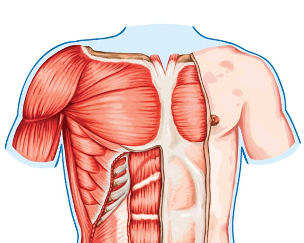
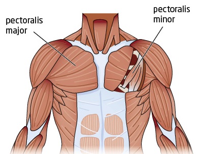
8 best upper chest exercises. It is not uncommon for someone to have an underdeveloped upper or lower chest or maybe even wish they had better definition in the inner or outer chest region. Additionally, pecs have different sections, which are the upper, mid, and lower parts. Atlas of anatomy of the human body: Find out more about the individual muscles within the chest the chest is part of a larger group of pushing muscles found in the upper body. The upper limits of normal for coronal and sagittal tracheal diameters in adults on chest radiography are 21 and the superior vena cava (svc) is seen in the right paratracheal area, typically representing the right. Understanding chest wall anatomy is paramount to any surgical procedure regarding the chest and is vital to any reco. Chest muscles affect movement of the arms and assist with shoulder function and stabilization. The embryologic and anatomic basis of modern surgery. Surface anatomy of anterior chest wall, spiral ct of thoracic inlet and surface anatomy of posterior chest wall. The chest anatomy includes the pectoralis major, pectoralis minor and the serratus anterior. Prime cuts bodybuilding dvds : Experts would obtain a preliminary supine scout radiograph of the chest with lead markers at 2cm intervals to localize the area of interest.